Research interests
Optical imaging is crucial for life science to study, understand and reveal new structures or phenomena. Recently, multi-cellular aggregates that mimics in vitro and in 3D some functions of organs (organoids) emerged as new biological models. Even if remarkable efforts have been made to adapt existing tools to image developing multicellular aggregates, current solutions are insufficient to observe the spatiotemporal organization of such samples over days or weeks in non-invasive manner.
My objective is to develop new imaging techniques or modalities dedicated to the non-invaisve observation of organoids.
Large-scale label-free microscopy
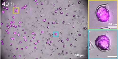
In the BiOf lab, we aim to develop simple, robust and cost-effective solutions to image large populations of multi-cellular aggregates (organoids) placed directly inside an incubator.
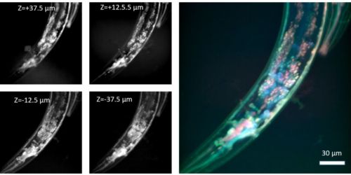
Multiplane confocal microscopy
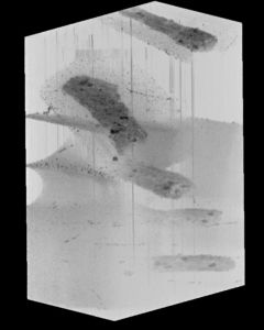
We recently developped an augmented variant of confocal microscopy where the key innovation consists to use a series of reflecting pinholes axially distributed in the detection plane, each one probing a different depth within the sample.
Light-field microscopy
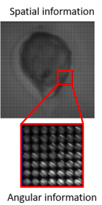
Light-field microscopy (LFM) offers instantaneous 3D imaging by capturing both spatial and angular information from a semi-transparent specimen. We aim to push back the current limits of LFM both in terms of depth and imaging volumes by combining innovative experimental and numerical approaches.
Supervision
Postdoctoral scientists :
- Naveen Mekhileri
PhD students :
- Aymerick Bazin (100 %)
- Léon Rembotte (10 %)
- Anirban Jana (20 %)
- Lucas Suire (50 %)
Selected publications
- A. Badon , S. Bensussen, H. J. Gritton, M. R. Awal, C. V. Gabel, X. Han and J. Mertz.
Video-rate large scale imaging with Multi-Z confocal microscopy, Optica 6 (4), 389-395
- G. Recher, P. Nassoy, A. Badon ,
Remote scanning for ultra-large field of view in wide-field microscopy and full-field OCT , Biomedical Optics Express 11 (5), 2578-2590
- A. Badon , V. Barolle, G. Lerosey, A. C. Boccara, M. Fink, A. Aubry.
Distortion matrix concept for deep imaging in scattering media, Science Advances 6 (30), eaay7170
- A. Badon , L. Andrique, A. Mombereau, L. Rivet, A. Boyreau, P. Nassoy, G. Recher.
The Incubascope: a simple, compact and large field of view microscope for long-term imaging inside an incubator, Royal Society Open Science 9 (2)
- A. Badon , G. Lerosey, A. C. Boccara, M. Fink, A. Aubry.
Retrieving time-dependent Green’s functions in optics with low-coherence interferometry, Physical Review Letters 114 (2), 023901 (IF 8.839)
