Principle
Light Field Microscopy (LFM) is a method that allows to simultaneously capture a large number of perspectives of the sample without any optical sectioning and then to digitally reconstruct the scene in 3D. This method is instantaneous, experimentally simple, but is still limited to small field of view, for instrumental reasons, and to shallow depths, due to the lack of true optical sectioning and for computational reasons, by analogy with how our brain would reconstruct the scene.
The aim of this project is to overcome the current limitations of LFM in terms of penetration depth, imaging modalities and field of view. The resulting imaging method will facilitate the non-invasive monitoring of large biological systems and decipher the propagation of light in complex scattering media. This project received fundings from the ANR JCJC Lifimmorg and the GPR Light.

Results
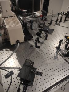
Currently our efforts are focused on the development of an experimental device allowing the capture of plenoptic images that will cover the entire field of view of the microscope objective without having to move the sample.
People involved in the project

Amaury Badon

Aymerick Bazin
Publications
Badon, A., Marceau, J. B., Allard, C., Fossard, F., Loiseau, A., Cognet, L., … & Gaufrès, E. (2023). Fluorescence Anisotropy Using Highly Polarized Emitting Dyes Confined inside BNNTs. Materials Horizons.